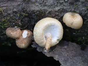
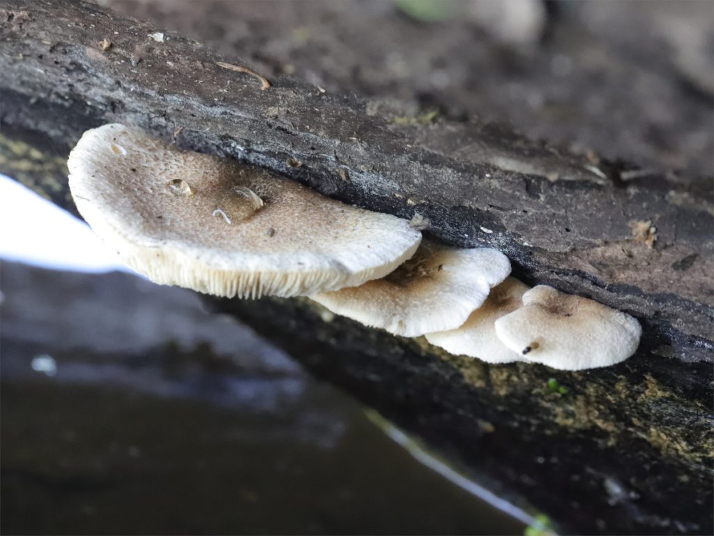
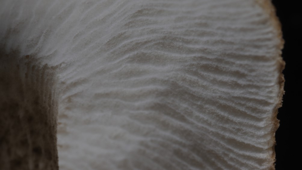
If you’ve read any of our other posts, you probably know by now that two distinct morphologies – agaricoid and secotioid – exist within the species Lentinus tigrinus. One of the first questions that follows is, “What’s causing it?” A major part of The Lentinus Project will be focused on answering that question.
In 1968, Rosinski and Robinson discovered that the agaricoid and secotioid forms of Lentinus tigrinus are mating compatible.1 This was an important insight because it settled the debate on whether the two forms were belong to a single species or represented two different species. The debate had been continuing slowly for decades, with good reason; the two forms are wildly different – I know of no other mushrooms where such a dramatic morphological difference exists in a single species. [I suppose you can get some weird shapes when mushrooms are infected with viruses, but that doesn’t really count.]

Further work by Rosinski and Faro (1968) and Hibbett et al. (1994) discovered that the two forms appear to be controlled by a single gene, with the agaricoid allele (version of a gene; aga) dominant over the secotioid allele (sec).2,3 Like humans, L. tigrinus has two copies of every gene – one copy inherited from each parent. If the sec allele is recessive, you would need two copies of the allele (sec/sec) to show the secotioid morphology. If you have two copies of the agaricoid allele (aga/aga) or one copy of each (aga/sec), the mushrooms would be agaricoid. We can therefore test whether a single gene is involved by crossing two individuals together and checking whether the morphologies of the children match our expectations (this is Mendelian genetics – you may remember doing Punnett squares in Biology class). When you do this work for L. tigrinus, the offspring match the predictions very well, so we assume the secotioid form is caused by a mutation in a single gene.

However, it’s never quite that straightforward in biology. There are many factors stored in DNA beyond just genes, so it’s not necessarily a gene that is responsible for the different morphologies. Genes are activated by nearby sections of DNA including promoters and regulators – the mutation could be to one of those sites. Or, perhaps there are several mutations in a group of genes so close together they behave as a single gene (a “supergene” – fungal mating loci are an example). Because we know a single region of DNA is responsible for the morphological difference but we don’t know what’s there, we will refer to this region as the “secotioid locus.”
Question
What is the nature of the secotioid locus?
Methods
Our lab has previously tried to identify the the secotioid locus, but with limited success. Work by Wu et al. in 2018 took an agaricoid mushroom and a secotioid mushroom and bred them together for three generations before sequencing a pool of 52 aga spores and a second pool of 49 sec spores.4 Statistical tests then identified 212 potential genes which could be the secotioid locus.
Because genomes were pooled together in the previous work, the contributions of individual genomes could not be assessed, which likely hampered the analysis. Our current project will sequence individuals separately so we can better resolve the secotioid locus during analysis. We will do this using two approaches: GWAS and population genetics.
GWAS
The primary method we will use to resolve the secotioid locus is Genome-Wide Association Studies (GWAS). For this technique, we will cross an aga/sec (agaricoid) individual with a sec/sec (secotioid) individual, fruit the children to identify them as either agaricoid or secotioid, and then sequence the genomes of 100 children. Bioinformatics software will then be used to determine which area(s) of the genome correspond to the secotioid morphology.
Population Genetics
For other parts of the project, we will be generating genomes from a population in Chicago, IL, and a population in Topsfield, MA. We can perform a similar analysis on these genomes to correlate morphology to DNA regions. Because there will naturally be more genetic variation in wild populations, this analysis will be less sensitive than the GWAS, but would allow us to double check the GWAS results using an independent dataset.
Outcomes
These analyses should allow us to narrow down the secotioid locus to a single genetic region comprised of a small number of genes and/or regulatory elements. Once we have identified the secotioid locus, we will be able to address more questions, such as: “How did the secotioid form evolve?” and “How does the locus alter development to create the secotioid form?”

References
- Rosinski, M. A. & Robinson, A. D. Hybridization of Panus Tigrinus and Lentodium Squamulosum. Am. J. Bot. 55, 242–246 (1968).
- Rosinski, M. A. & Faro, S. The genetic basis of hymenophore morphology in Panus tigrinus. (Bull. ex Fr.) Singer. Am. J. Bot. 55, 720 (1968).
- Hibbett, D. S., Tsuneda, A. & Murakami, S. The secotioid form of Lentinus tigrinus: genetics and development of a fungal morphological innovation. Am. J. Bot. 81, 466–478 (1994).
- Wu, B. et al. Genomics and Development of Lentinus tigrinus: A White-Rot Wood-Decaying Mushroom with Dimorphic Fruiting Bodies. Genome Biol. Evol. 10, 3250–3261 (2018).

